X-ray Diffraction Microscopy
Lensless Microscopy using Coherent X-rays
X-rays has a short wavelength of ~0.1 nm, and thus can overcome the resolution limitation ~200 nm in conventional optical microscopy. In addition, X-ray has a high penetration power, and thus allows us to observe structures deep inside an object, which are difficult to access with electron microscopy. However, since x-rays can transmit even a thick human body, it passes through micrometer-sized cells with little interaction. As it is difficult to observe air with our eyes, ingenuity is required to observe “transparent” cells.
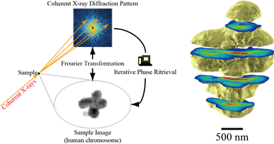
Coherent x-rays with a beautiful wave pattern plays an important role in observing transparent objects, and is available at advanced synchrotron radiation facilities, such as SPring-8.
Fig. Observation of Human Mitotic Chromosome in 2D & 3D by X-ray Diffraction Microscopy
References
- Three-Dimensional Visualization of a Human Chromosome Using Coherent X-Ray Diffraction
Yoshinori Nishino, Yukio Takahashi, Naoko Imamoto, Tetsuya Ishikawa, and Kazuhiro Maeshima
Physical Review Letters 102, 018101 (2009).
<color red>World's first 3D bioimaging by x-ray diffraction microscopy</color> - Imaging Whole Escherichia Coli Bacteria by Using Single-Particle X-Ray Diffraction
Jianwei Miao, Keith O. Hodgson, Tetsuya Ishikawa, Carolyn A. Larabell, Mark A. LeGros, and Yoshinori Nishino
Proceedings of the National Academy of Sciences of the United States of America, 100, 110-112 (2003).
<color red>World's first 2D bioimaging by x-ray diffraction microscopy</color>

